Decoding Cell Surface Sugars: A New Frontier for Early Cancer Detection
Every human cell is coated with a complex layer of sugar molecules that form unique patterns—a kind of molecular fingerprint. Scientists from the Max Planck Institute have developed an advanced imaging technique called Glycan Atlasing to visualize these sugar structures. They discovered that these patterns change when cells are activated or become cancerous. This breakthrough offers a potential new way to detect cancer early by spotting telltale sugar signatures on cell surfaces. Below we explore the key findings and implications of this research.
What is the "sugar code" on human cells and why is it important?
The "sugar code" refers to the intricate arrangement of sugar molecules, known as glycans, that coat the outer membrane of every human cell. These glycans form specific patterns that vary depending on the cell type, its state, and its interactions with the environment. Think of them as a molecular ID card that cells use to communicate and identify each other. The sugar code is crucial because it influences how cells recognize signals, adhere to surfaces, and interact with the immune system. Changes in these patterns can indicate a shift in cellular behavior—such as activation, stress, or transformation into a cancerous state. By reading this code, scientists hope to gain early insights into disease processes long before symptoms appear, making it a promising target for next-generation diagnostics and therapies.
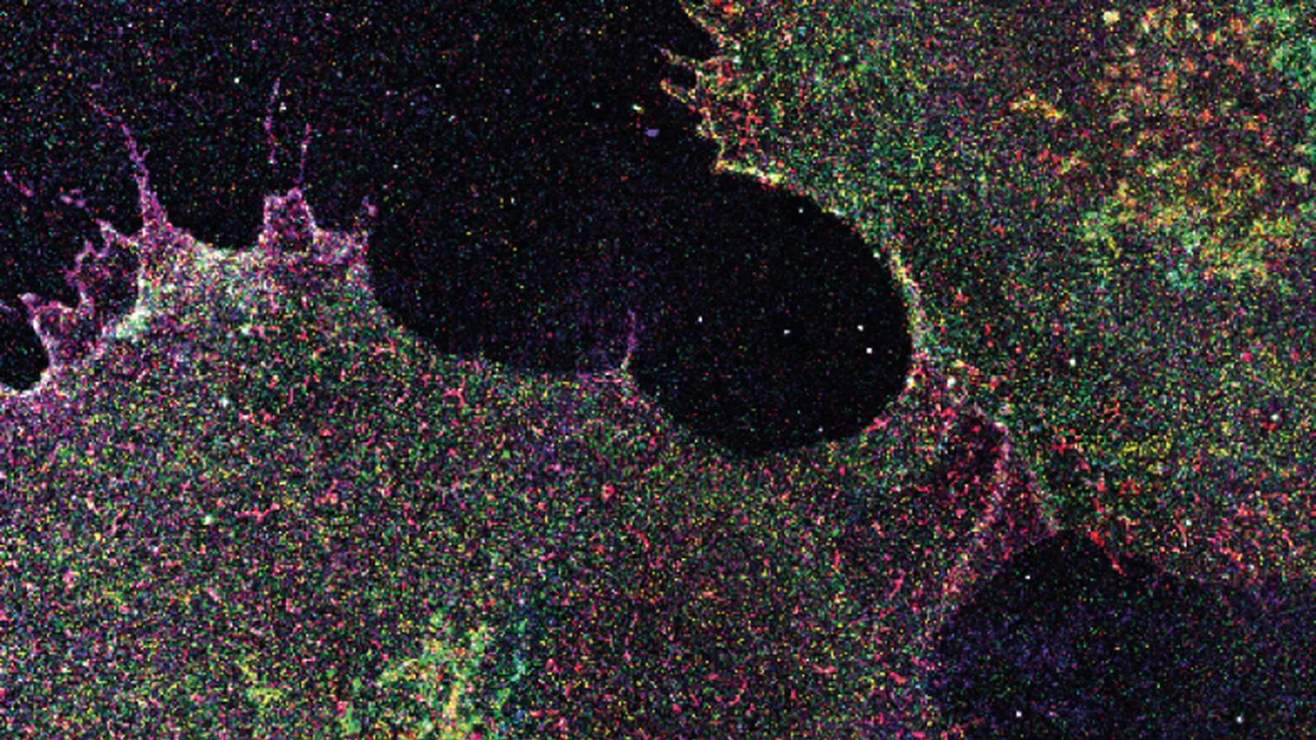
How did researchers map these sugar patterns?
To decode the sugar patterns, researchers at the Max Planck Institute developed a cutting-edge technique called Glycan Atlasing. This method combines high-resolution imaging with advanced data analysis to create detailed maps of glycans on cell surfaces. The team used specialized probes that bind to specific sugar structures, allowing them to visualize the distribution and density of glycans with unprecedented clarity. By analyzing these maps from thousands of individual cells, they could identify consistent patterns and variations. The technique enabled them to compare sugar layouts across different cell types, states, and conditions—including healthy vs. cancerous tissues. Glycan Atlasing effectively turns the invisible sugar code into a visible, quantifiable dataset, opening the door to systematic study of how these patterns relate to health and disease.
How do sugar patterns change in immune cells when activated?
When immune cells are activated—for instance, in response to an infection—they undergo dramatic changes in their surface sugar layouts. The researchers observed that resting immune cells have a relatively simple and uniform glycan coat. However, once activated, the cells produce a more diverse and complex set of sugars. These changes are not random; they follow a specific program that helps the immune cells migrate to sites of inflammation, recognize pathogens, and communicate with other immune cells. For example, certain sugars become more abundant to facilitate cell-to-cell adhesion, while others are modified to alter signaling pathways. This dynamic remodeling of the sugar code is essential for a proper immune response. By tracking these shifts, scientists can better understand how immune cells function and how they might go awry in autoimmune diseases or chronic inflammation.
What differences were found between cancerous and healthy tissues?
Comparing cancerous tissues to healthy counterparts, the researchers discovered distinct and reproducible differences in the sugar patterns. Cancer cells consistently displayed a unique fingerprint of glycans on their surface that differed from normal cells in both composition and arrangement. Notably, many cancer cells showed an increased presence of specific sugar chain lengths or branching patterns, as well as altered expression of certain sugar molecules. These changes are thought to help cancer cells evade immune detection, promote metastasis by adhering more easily to blood vessels, and drive uncontrolled growth. Importantly, these sugar signatures were found even in early-stage tumors, before they become large or invasive. This suggests that cancer-related glycan alterations occur early in the disease process, making them ideal candidates for early detection tests that could identify cancer from a simple blood sample or biopsy.
How could this discovery lead to early cancer detection?
The discovery that cancerous tissues have distinct, early-emerging sugar patterns opens the door to non-invasive screening methods. Because these glycan signatures are present on the surface of cancer cells, they can potentially be detected in bodily fluids—such as blood, urine, or saliva—when tiny fragments of tumor cells or their secreted glycans circulate. Researchers envision developing diagnostic tests that use specific probes or antibodies to recognize and quantify these cancer-associated sugar markers. Such tests could detect cancer at its earliest, most treatable stage, before tumors are visible on imaging scans or cause any symptoms. Moreover, the technique could be used to monitor treatment response, as changes in sugar patterns might indicate whether a therapy is working. While still in research stages, the potential for a simple, cost-effective, and highly sensitive early cancer detection tool based on the sugar code is extremely promising.
What are the next steps for this research?
Following this initial mapping of glycan patterns, the Max Planck team plans to expand their studies to a wider range of cancer types and stages. They aim to build a comprehensive atlas of sugar signatures associated with different cancers, including breast, lung, colon, and pancreatic cancers. Next steps also involve developing highly specific probes and assays that can detect these sugar changes in clinical samples with high accuracy. Collaborations with medical centers will be essential to validate the findings in patient biopsies and blood samples. Additionally, researchers will investigate how the sugar code changes over the course of cancer progression and treatment. Ultimately, the goal is to translate these fundamental discoveries into practical diagnostic tools that can be deployed in hospitals and clinics. The journey from lab to clinic may take several years, but the sugar code is emerging as a powerful new biomarker for early cancer detection.